
Stent Thrombosis Risk Calculator
Stent Thrombosis Risk Assessment
This tool calculates your personalized risk of stent thrombosis based on stent type and clinical factors. Results are for informational purposes only.
Your Results
DAPT Duration Recommendation:
Notes: Risk assessment is based on clinical guidelines. Your physician will consider all factors for personalized treatment.
Key Takeaways
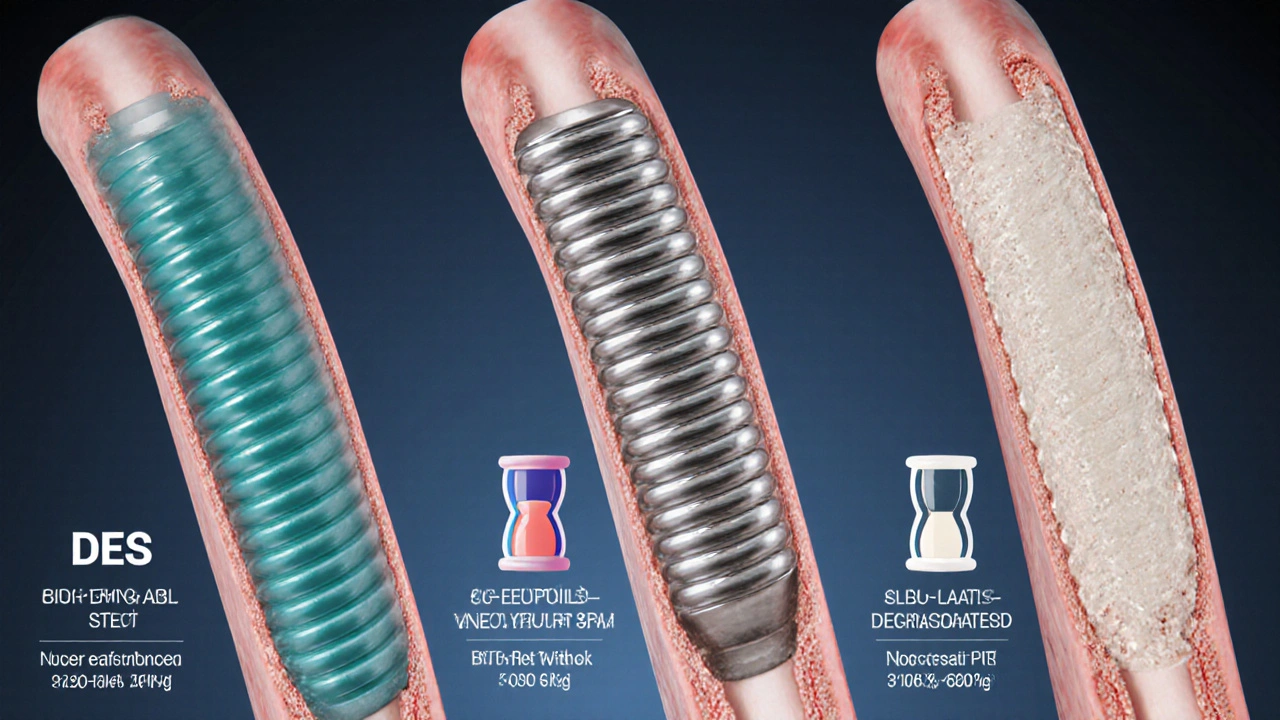
- Stent thrombosis is a rare but life‑threatening complication that varies by stent design.
- Drug‑eluting stents (DES) lower restenosis but need longer dual antiplatelet therapy (DAPT).
- Bare‑metal stents (BMS) carry a higher restenosis risk yet allow shorter DAPT.
- Bioresorbable scaffolds offer a temporary framework but have specific implantation rules.
- Patient factors (diabetes, smoking, poor adherence) dramatically influence clot risk.
When a stent fails because a clot forms inside it, doctors call the event stent thrombosis is the acute blockage of a coronary artery by a blood clot forming on or within a previously placed stent. This complication can cause a heart attack, sudden cardiac death, or need for emergency re‑intervention. Understanding why it happens-and how different stent designs affect the odds-helps patients and clinicians make smarter choices after a percutaneous coronary intervention (PCI).
Below we break down the main stent families, their engineering trade‑offs, and the clinical steps that keep clot formation at bay.
What’s Inside a Stent? Core Components
All coronary stents share three basic parts:
- **Scaffold material** - usually stainless steel, cobalt‑chromium, or a polymer that dissolves over time.
- **Drug coating** - for DES, a thin layer releases medication that thwarts smooth‑muscle cell overgrowth.
- **Strut geometry** - thickness and pattern that affect blood flow and endothelial healing.
These elements dictate how quickly the artery re‑endothelializes (the natural healing that lines the vessel) and how long patients must stay on antiplatelet drugs.
Types of Stents and Their Thrombosis Profiles
Each stent type carries a distinct clot risk, shaped by material, drug, and design.
Drug‑eluting stent (DES) is a metallic scaffold coated with an antiproliferative drug (e.g., everolimus, zotarolimus) that slowly releases into the artery wall to prevent restenosis. Modern DES have ultra‑thin struts (60‑80µm) and biodegradable polymers that reduce inflammation.
- Thrombosis risk: Early‑generation DES (first‑gen) showed higher late‑stage clot rates; newer DES (second‑gen and beyond) have <0.5% annual stent thrombosis.
- DAPT duration: Typically 6-12months, longer if high bleeding risk is not a concern.
- Restenosis rate: 5‑7% versus 20‑30% for BMS.
Bare‑metal stent (BMS) is a simple metallic scaffold without any drug coating. BMS were the first devices used in PCI and remain in use where short‑term antiplatelet therapy is needed.
- Thrombosis risk: Similar to early DES in the first months, but slightly higher after 1year if endothelialization is incomplete.
- DAPT duration: Usually 1-3months, making BMS attractive for patients with high bleeding risk.
- Restenosis rate: 20‑30%-the main drawback.
Bioresorbable vascular scaffold (BVS) is a temporary, polymer‑based stent that fully dissolves over 2-3years, leaving a natural artery behind. The concept promises no permanent metal, potentially reducing late‑stage thrombosis.
- Thrombosis risk: Early trials reported higher early‑stage clot rates (up to 2%) due to thicker struts and suboptimal lesion preparation.
- DAPT duration: Often 12months or more, plus strict implantation technique (PSP: pre‑dilation, sizing, post‑dilation).
- Restenosis rate: Comparable to contemporary DES when deployed correctly.
Why Do Clots Form? The Physiology Behind Stent Thrombosis
Three mechanisms usually converge:
- Platelet activation: Stent struts disturb laminar flow, exposing subendothelial collagen and triggering platelets.
- Incomplete endothelialization: If the vessel lining doesn’t fully cover the stent, blood contacts foreign material, keeping the clotting cascade active.
- Inflammatory response: Polymer residues or metal ions can provoke local inflammation, encouraging thrombus formation.
Patient‑specific factors add another layer of risk. Diabetes, chronic kidney disease, smoking, and a history of acute coronary syndrome all raise the odds of clotting.
Antiplatelet Strategies: Keeping the Blood Flowing
Dual antiplatelet therapy (DAPT) is the combination of aspirin and a P2Y12 inhibitor (clopidogrel, prasugrel, or ticagrelor) used to blunt platelet aggregation after stent placement. The balance between clot prevention and bleeding risk drives the chosen duration.
- Standard DAPT: 6months for most DES, 1month for BMS, 12months for BVS.
- Short‑DAPT strategies: In high‑bleeding‑risk patients, 1‑month DAPT with a newer DES (e.g., polymer‑free) is becoming acceptable.
- Extended DAPT: For patients with prior myocardial infarction or complex lesions, 24‑month DAPT can further lower late thrombosis.
Adherence matters. Missed doses of the P2Y12 blocker dramatically increase early stent thrombosis risk-studies show a three‑fold rise within the first 30days.
Comparing Stent Types - Quick Reference
| Attribute | Drug‑Eluting Stent (DES) | Bare‑Metal Stent (BMS) | Bioresorbable Scaffold (BVS) |
|---|---|---|---|
| Material | Cobalt‑Chromium or Platinum‑Chromium | Stainless Steel or Cobalt‑Chromium | Poly‑L‑lactic acid (PLLA) polymer |
| Drug coating | Everolimus, Zotarolimus, Sirolimus | None | None (structural polymer only) |
| Typical DAPT length | 6-12months (up to 24months for high risk) | 1-3months | 12months (or longer if needed) |
| Restenosis rate | 5‑7% | 20‑30% | 5‑10% (when properly implanted) |
| Late stent thrombosis (<1year) | 0.2‑0.5% | 0.5‑1% | 1‑2% |

Practical Steps to Lower Your Clot Risk
- Follow DAPT instructions to the letter. Set alarms, use pill organizers, and keep regular follow‑up appointments.
- Control modifiable risk factors. Quit smoking, keep blood sugar under control, and maintain a healthy lipid profile.
- Know your stent type. Ask your interventional cardiologist which stent was used and the recommended DAPT duration.
- Report symptoms early. Chest pain, shortness of breath, or sudden fatigue may signal a clot; seek emergency care.
- Stay informed about newer guidelines. Trials like STOPDAPT‑2 and TWILIGHT have reshaped DAPT recommendations for specific groups.
When to Consider a Switch in Therapy
If a patient experiences bleeding complications or cannot tolerate clopidogrel, clinicians may switch to a newer P2Y12 inhibitor (prasugrel or ticagrelor) or use a single‑antiplatelet strategy after the minimum DAPT period. Recent data suggest that, in selected low‑risk patients with newer DES, stopping aspirin while continuing a P2Y12 blocker can keep thrombosis rates low while cutting bleeding risk.
Future Directions - What’s on the Horizon?
Researchers are testing ultra‑thin strut DES with fully bio‑absorbable polymers, aiming for <0.2% late thrombosis without extending DAPT. Parallel work on surface‑engineered BMS (e.g., nano‑textured coatings) hopes to speed endothelialization, making short DAPT safe for a broader population.
Frequently Asked Questions
What is the difference between early and late stent thrombosis?
Early thrombosis occurs within 30days of implantation and is usually linked to procedural issues, poor stent expansion, or premature discontinuation of antiplatelet drugs. Late thrombosis happens after 30days, often driven by delayed endothelialization, chronic inflammation from polymer residues, or patient‑related factors like diabetes.
Can I stop aspirin early if I’m on a drug‑eluting stent?
Recent trials (e.g., TWILIGHT) suggest that after 3months of DAPT with a newer DES, stopping aspirin while continuing ticagrelor may reduce bleeding without raising clot risk. However, this approach should only be taken under cardiologist supervision and is not standard for all patients.
Are bioresorbable scaffolds safer than metal stents?
Bioresorbable scaffolds remove the permanent metal cage, potentially lowering very late thrombosis. Yet, early‑generation BVS showed higher early clot rates because of thicker struts. Newer designs with thinner struts are narrowing the safety gap, but long‑term data are still limited.
How does diabetes increase stent thrombosis risk?
Diabetes promotes platelet hyper‑reactivity, endothelial dysfunction, and slower healing of the vessel wall. These factors together raise both early and late thrombosis rates, making strict glycemic control and adherence to DAPT crucial.
What imaging tools help detect early stent thrombosis?
Coronary angiography remains the first line, but intravascular ultrasound (IVUS) and optical coherence tomography (OCT) can visualize strut coverage and detect thrombus that angiography might miss.


Henry Seaton
Stent choices shouldn’t be taken lightly. The medical system expects us to follow the guidelines not play roulette with lives.
Baby Thingie
Your risk calculator correctly emphasizes dual antiplatelet therapy duration. :)
Abby Elizabeth
omg i cant even with all this stent drama lol. like why does every doc act like they’re the only one who knows? i feel sooo lost.
Barbra Wittman
Oh, because we all love a good risk calculator that pretends to replace a cardiologist’s judgment.
It’s adorable how the interface asks you to pick a stent type like you’re ordering pizza.
Do you also want extra cheese with your drug‑eluting stent?
And let’s not forget the overly bright risk‑badge colors that scream “look at me, I’m dangerous!”
Sure, the tool tells you the DAPT duration, but it conveniently omits the fact that adherence is a personal habit, not a button you can click.
Speaking of habits, did anyone notice that the “non‑adherence” checkbox is hidden behind a tiny toggle?
It’s almost as if the designers assume we’ll never remember to check it.
Maybe the next version will include a therapist to coach you through quitting smoking while you wait for the stent to dissolve.
Because, of course, a bioresorbable scaffold solves all of our problems, right?
In reality, those scaffolds have their own set of strict implantation rules that most interventionalists find cumbersome.
And yet the calculator pretends they’re as simple as a BMS.
Let’s also applaud the bold claim that “risk assessment is based on clinical guidelines” while ignoring patient‑specific nuances.
If you’re a clinician, you’ll know we already have guidelines; we don’t need a flashy web page to remind us.
But for the layperson, this tool might feel like a magic eight ball that spits out risk levels with no context.
Bottom line: use it as a conversation starter with your doctor, not as a definitive diagnosis.
Lynnett Winget
Hey, great job breaking down the stent families – the colorful analogies really help me picture the differences.
Just remember, every patient’s story adds another shade to the picture, so keep the dialogue open.
Amy Hamilton
The balance between preventing restenosis and minimizing thrombosis risk is a classic philosophical dilemma in interventional cardiology.
Choosing a newer DES with a biodegradable polymer reflects a thoughtful compromise, acknowledging both science and patient lifestyle.
In practice, aligning the DAPT duration with individual risk factors exemplifies personalized medicine at its best.
Lewis Lambert
If you’re comfortable with a short DAPT regimen, a BMS can be a pragmatic choice, especially when adherence is a concern.
Just keep a close eye on any signs of restenosis during follow‑up.
Jordan Schwartz
I appreciate the balanced tone here – it’s reassuring for anyone navigating these decisions.
Remember, open communication with your cardiologist can turn these guidelines into a tailored plan.
Nitin Chauhan
Stay motivated to stick with your medication schedule; consistency beats occasional bursts every time.
Even without emojis the message is clear – keep pushing forward.
Dannii Willis
Interesting overview – the mix of formal data and friendly tone makes it accessible.
Looking forward to seeing more on long‑term outcomes.
Robyn Du Plooy
From a hemodynamic perspective, the strut thickness directly influences shear stress gradients, which in turn modulate endothelialisation kinetics.
Thus, ultra‑thin DES platforms often exhibit reduced thrombotic propensity compared to bulkier BMS designs.
Boyd Mardis
Pick the stent that aligns with your lifestyle and stick to the prescribed DAPT schedule.
Zach Yeager
Some folks think the calculator replaces professional advice but it really just adds a layer of confusion.
Better to trust the clinic over a website.
Angel Gallegos
The presentation attempts sophistication yet falls short of depth; the analysis feels superficial at best.
Consider a more rigorous review before drawing conclusions.